Texbook: Ganong's Review of Medical Physiology 27th Edition, Chapter 29 + page 122-126
% of MCQ: 25% 'The Heart and Circulation'
Electrical Activity of the Heart
- Cardiac excitation
- The electrocardiogram
- Cardiac arrhythmias
- Electrocardiographic findings in other diseases
Cardiac muscle cells
Properties:
- Involuntary
- Striations, Z lines, branching cells, T tubules
- One central nucleus
- Intercalated discs connecting myocytes via gap junctions, allowing rapid electrical spread
- Abundant mitochondria
- High myoglobin content (stores O2)
- Limited regenerative capacity, meaning injury tends to lead to fibrosis
Functional types: Pacemaker cells (AV/SA node), conducting cells (His-Purkinje) and contractile myocytes
Metabolism: aerobic (high O2 requirements)
Chain of events: SA node generates action potential-->electrical signal passes between myocytes via gap junctions-->signal penetrates through cell membrane (sarcolemma) and goes down deep membrane invaginations known as T-tubules to come adjacent to the sarcoplasmic reticulum(SR)--> L-type Ca²⁺ channels open and Ca²⁺ enters-->RYR2 on SR activated and Ca²⁺ is released from SR -->Ca²⁺ binds Troponin C-->conformational change--> Troponin I mediated inhibition is relieved->Tropomyosin, which is associated with the troponin complex via Troponin T, moves away from the myosin-binding sites on actin->Actin and myosin cross-bridge cycling-->contraction-->L-type Ca²⁺ channels close-->SERCA puts Ca²⁺ back into SR, Na/Ca exchanger pumps Ca²⁺ out-->Ca²⁺ leaves troponin C,-->Troponin I mediated inhibition is restored-->Tropomyosin moves back over the myosin-binding sites on actin-->relaxation
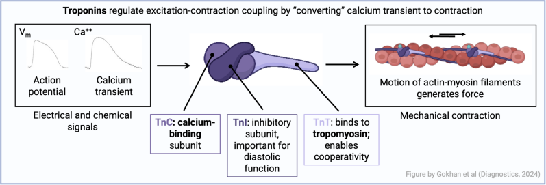
Gokhan I, Dong W, Grubman D, Mezue K, Yang D, Wang Y, Gandhi PU, Kwan JM, Hu J-R. Clinical Biochemistry of Serum Troponin. Diagnostics. 2024; 14(4):378. https://doi.org/10.3390/diagnostics14040378
Action potentials
Depolarisation: when inside the cell becomes LESS negative, and thus less polarised (i.e. + ions move IN)
Repolarisation: when inside the cell becomes MORE negative again, returning to its resting potential (i.e. + ions move OUT)
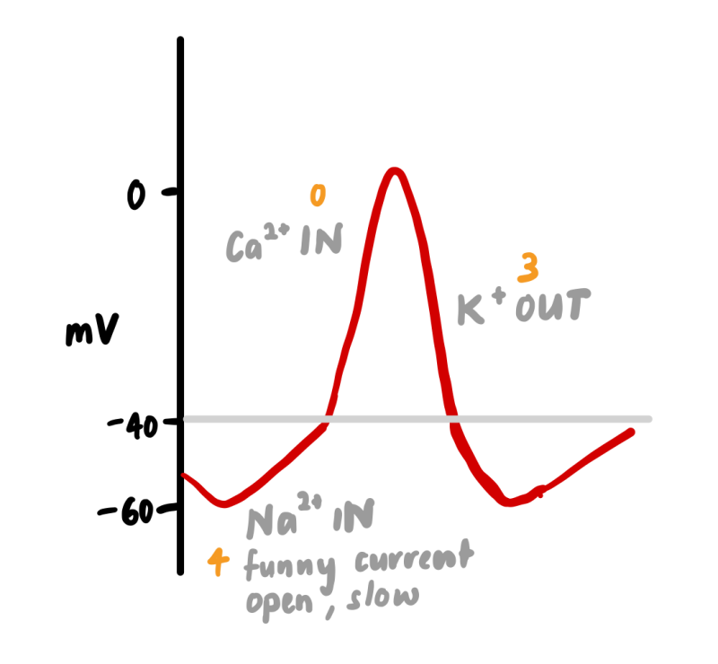
Draw an action potential for a pacemaker cell
Pacemaker cells are modified myocytes. The SA node is the primary pacemaker of the heart. The repeating change in membrane voltage of cardiac pacemaker cells allows them to spontaneously generate impulses that travel to contractile myocytes via gap junctions. They have no true 'resting membrane potential'.
Phase 0 = Ca²⁺ influx
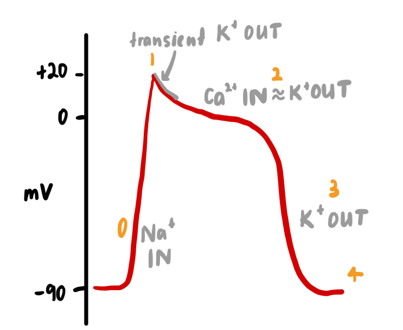
Draw an action potential for a ventricular myocyte
Resting membrane potential= -90mV
Phase 0 = Na⁺ influx
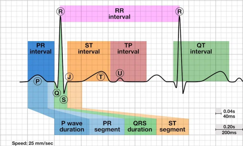
ECG fundamentals
P wave: atrial depolarisation. Peaked P waves ("P pulmonale") indicates right atrial enlargement. Bifid P waves ("P mitrale") indicates left atrial enlargement. Upright P waves in lead I and II with negative in avR (inverted) is what defines "sinus" rhythm i.e. rhythm coming from sinus node. Negative in II or positive in aVR suggests low atrial/junctional origin (or lead error).
Q wave: the first negative deflection. It is normal to have small septal q waves (due to normal L>R septal depolarisation) and sometimes seen in inferior leads, but should be narrow and small. Abnormally wide (>40ms) or deep (>2mm) or >25% of the following R wave amplitude is pathological and usually mean infarction or scar tissue. Note HOCM can produce deep, but narrow 'dagger-like' Q waves.
R wave: the first positive deflection of the QRS complex. It reflects early ventricular depolarisation, predominantly of the left ventricle. R-wave amplitude should increase from V1 to V6, with R ≈ S around V3-V4 normally.
- Early transition (R ≈ S≤V2): Rightward rotation, RVH, WPW type A, posterior OMI reperfusion, or lead misplacement (V1–V2 too low or too lateral).
- Late transition (R ≈ S≥V5): Leftward rotation, LVH, LBBB, prior anterior MI, COPD hyperinflation, or high placement of V1–V2.
- Posterior OMI pattern: Tall R in V1–V2 plus ST depression V1–V3 is ischaemic until proven otherwise.
S wave: first negative deflection after the R wave within the QRS. Normally deep in V1–V2, shallower across V3–V6.
- Dominant S in V5–V6 with tall R in V1 can mean RVH or RBBB with RVH.
- Deep S in V1–V3 can mean LVH.
- S1Q3T3 pattern: prominent S in I, Q in III, T inversion in III with rightward axis or RBBB features. Use only in the right clinical context.
T wave: reflects ventricular repolarisation. Normally upright in I, II, V3–V6, negative in aVR and variable in III, aVL, V1–V2. New inversion can mean ischaemia. Peaked in hyperkalaemia, flat in hypokalaemia, biphasic in Wellens. de Winter T waves are an OMI pattern, suggesting proximal LAD occlusion.
AXIS
- Positive electrical movement towards the lead= upward deflection
- Positive electrical movement away from the lead= downward deflection
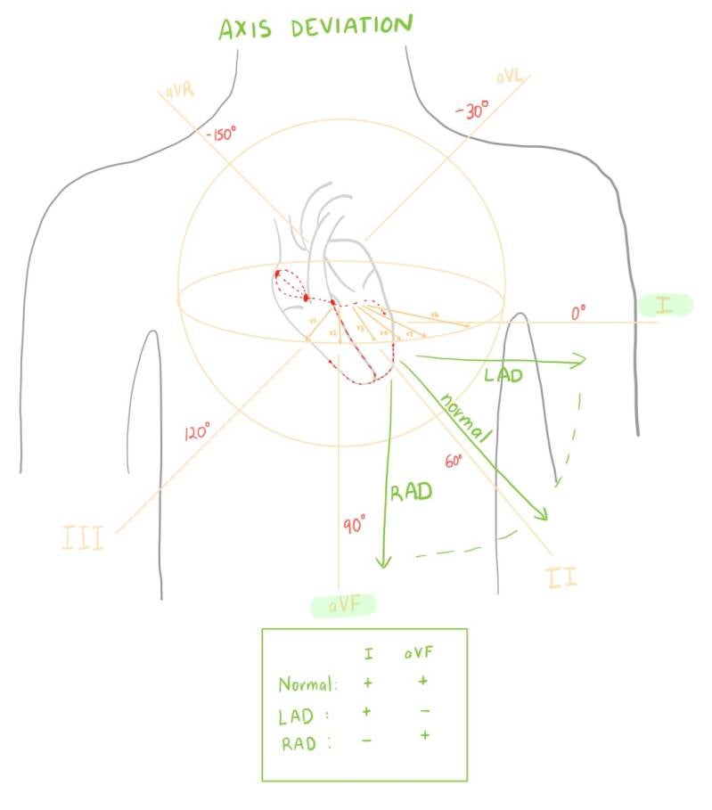
Describe the spread of cardiac excitation with reference to relative conduction speeds (and thus the normal intervals on ECG)
PR interval
Events in the heart: Atrial depolarisation plus conduction through the AV node and His-Purkinje system. Reflects the time between atrial activation and ventricular activation. Normal AV delay is 0.1s.
Normal duration: 0.12-0.2ms
Prolonged PR (>200 ms)= first-degree AV block. Drugs that prolong AV node conduction: beta blockers, diltiazem, verapamil, digoxin, amiodarone. Also prolonged in sleep (high vagal tone).
Short PR (<120 ms)= think about pre-excitation (if delta wave present, with WPW)
QRS
Events in the heart: Ventricular depolarisation
Normal duration: 0.06-0.1 ms; Borderline 0.1-0.12ms; Wide >120ms (3 small squares)
Widened=BBB, VT/VF, sodium channel blocker toxicity(e.g. TCAs), hyperkalaemia, WPW (+ delta wave), pacemaker.
Note that if the QRS is widened in one lead, it should be the same in every lead. Sometimes a widened QRS can look like ST elevation, so it is useful to compare its width to the other leads.
Narrow=normal, SVT.
QT
Events in the heart: Ventricular action potential
Normal duration: < 440-450 milliseconds (ms) for men and <460-470 ms for women.
Ensure you correct for HR:
Fridericia: QTc = QT / ∛RR (more accurate with significant tachy/brady cardia)
Bazett: QTc = QT / √RR (most common)
Risk of Torsades rises substantially when QTc ≥500 ms.
Long QTc=Hypocalcaemia, Hypomagnesaemia, Hypokalaemia, Hypothermia, Medications, CNS bleed/injury e.g. SAH.
If the T wave ends after halfway to the next R wave, suspect QT prolongation.
Short QTc(<350 ms); consider hypercalcemia
What effect does a 'block' have on the spread of cardiac excitation
Junctional escape rhythm
Whilst it is not a 'block' as such, when the sinus node fails, you get a protective 'junctional' rhythm which takes over, arising from the AV junction (usually the AV node or proximal His bundle). This 'junctional escape rhythm' is usually 40-60bpm and hallmarked by P waves which are absent, inverted in the inferior leads, buried within the QRS, or appear after the QRS due to retrograde atrial activation. The QRS are typically narrow, but can be wide is there is a preexisting bundle branch block, aberrancy, or ventricular conduction disease.
AV Blocks
The SA node usually controls the heart rate, because its discharge rate is higher than other parts of the conduction system. Normal AV delay is 0.1s.
- AV block; 45bpm
- Infranodal block; 35bpm
1st degree
Long PR > 200 ms but 1:1 P:QRS conduction with no dropped beats
Usually benign, medications e.g. beta blockers
2nd degree Mobitz I (Wenchebach)
Progressive lengthening until dropped QRS.
Two features that quickly confirm Wencheback: 1) The post-drop PR is shorter than the last conducted PR and 2) the pause containing the block is < 2x P-P interval. The issue is usually in AV node. Low pacing need.
2nd degree Mobitz II
Fixed PR with intermittent dropped QRS
Issue usually infranodal (in His-prukinje system). Mobitz II is dangerous. Often needs PPM. This there is often also an existing bundle branch block.
2:1 AV block
Every other P wave conducts. PR may be constant or lengthening but you cant type it without two consecutive conducted beats.
Narrow QRS= nodal cause
Widened QRS= infranodal cause
3rd degree
P waves and QRS complexes follow independent rhythms without conducted beats. Regular escape rhythm present; junctional (narrow QRS, 40-60 bpm) or ventricular (wide QRS, 20-40bpm). Complete AV dissociation which requires PPM. Note that if you see ANY conducted beats, the block is high-grade second degree rather than complete. Can be caused by septal MI.
Bundle Branch Blocks
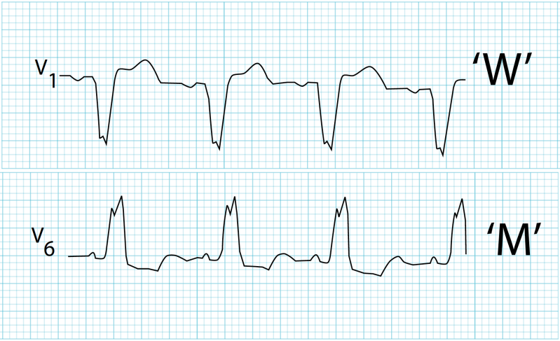
Left BBB
- Conduction spreads down right bundle to RV, then to the LV via the septum. Since the LV is bigger, the dominant vector is still negative in V1 (right side of heart), and positive in V6 (left side of heart). There is a 'W' appearance in V1 and 'M' appearance in V6.
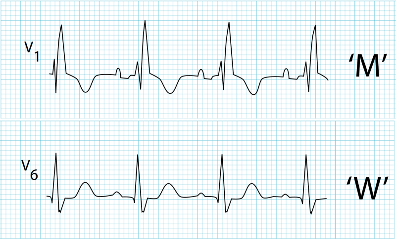
Right BBB
- Conduction spreads down left bundle to LV, then to the RV via the septum. The dominant vector is negative in V1, with a late positive deflection ('M' appearance), and positive in V6, with a late negative deflection ('W' appearance)
Fascicular blocks
Left anterior fascicular block
- Activation of inferior/posterior LV first, which then spread up and left
- Left Axis deviation with R wave peak time >45ms in aVL
- QRS < 120ms
Left posterior fascicular block (less common as posterior fascicle has dual blood supply)
- Activation of anterior LV first, then spreads down and right
- Right axis deviation with R wave peak time >45ms in aVF
Bi-fascicular block
- Widened QRS+ LAD = RBBB + LAFB = most common
- Widened QRS + RAD = RBBB + LPFB
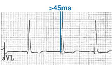
LAFB: https://litfl.com/left-anterior-fascicular-block-lafb-ecg-library/
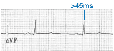
LPFB: https://litfl.com/left-posterior-fascicular-block-lpfb-ecg-library/
What causes the major abnormalities in the ECG in Acute MI
Note that almost any CHANGE to someone's ECG can represent ischaemia e.g. a new bundle branch block. Thus serial ECGs are an essential part of determining whether a patient is having an MI.
T wave: Ischaemia alters myocardial repolarisation, producing abnormal T-wave vectors.
- Early ischaemia can produce hyperacute, broad-based T waves. De Winter T waves= sign of LAD occlusion.
- Later, T-wave inversion may develop
ST segment: The ST segment is when the ventricular myocardium is essentially uniformly depolarised. So there is little voltage difference between different regions of the ventricle → little net electrical current → isoelectric 'flat' ST segment. Ischaemia initially increases the membrane potential of the infarcted area, creating a voltage difference (normal region is relatively negative compared to infarcted area).
- ST elevation → abnormal rapid repolarisation, decreased resting membrane potential, delayed depolarisation (due to accelerated opening of K+ channels)
- ST depression → injury vector directed away from the lead / sub-endocardial ischaemia pattern
- Reciprocal changes occur in electrically opposing leads
Q wave: abnormally wide (>40ms) or deep (>2mm) Q waves, or >25% of the following R wave amplitude is pathological and usually mean infarction or scar tissue. Note that HOCM can produce deep, but narrow 'dagger-like' Q waves.
| Area of infarct | Leads with ST elevation | Coronary Artery implicated |
|---|---|---|
| Inferior | II, III, aVF | RCA |
| Anterior | V3-V4 | LAD |
| Septal | V1-V2 | LAD |
| Lateral | I, aVL, V5-V6 | LAD/LCx |
| Posterior | V7-V9, reciprocal STD V1-V2 | LCx/RCA |
Benign early repolarisation: common pattern characterized by J-point elevation, often with concave ST elevation and J-wave notching or slurring. Most commonly seen in precordial and lateral leads (less common in inferior leads). No reciprocal ST depression. More common in young healthy adults, black men.
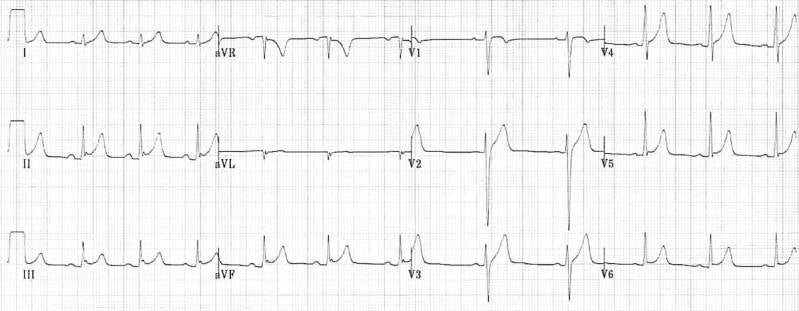
What is the effect of a changing K+ concentration on the ECG
Normally NA/K ATP-ase(3 Na⁺ out, 2 K⁺ in) maintains high K inside cell (low outside cell)
In hyperkalaemia: ↑ extracellular K⁺ → smaller K⁺ concentration gradient → K⁺ has less tendency to leave → membrane potential becomes less negative → initially altered repolarisation→peaked T waves first sign→if K >9 (severe) Na⁺ channel inactivation→ PR prolongation + QRS widening→"Sine-wave" ECG, VF
In Hypokalaemia(K<2.7): ↓ extracellular K⁺→ larger K⁺ concentration gradient→ alters K⁺ currents and ventricular repolarisation→ prolonged repolarisation→ T-wave flattening + U waves + prolonged QU
Other electrolytes:
- Hypercalcaemia; short QT
- Hypocalcaemia: long QT
- Low Na: smaller QRS complexes
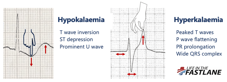
https://litfl.com/hyperkalaemia-ecg-library/
What is the effect of sympathetic stimulation on the electrical activity of the heart?
Sympathetic stimulation
In SA node (pacemaker cell)
- β₁-adrenoceptor agonism → Gs → ↑ adenylyl cyclase → ↑ cAMP → ↑ PKA →↑ If ("funny") current and Ca²⁺ currents → faster phase 4 depolarisation (pacemaker cells reach threshold more rapidly as resting membrane potential less negative) → ↑ heart rate
- Temperature and drugs also affect the rate of discharge of the SA node
In the AV node (pacemaker cell)
- β₁-adrenoceptor agonism → Gs → ↑ adenylyl cyclase → ↑ cAMP → ↑ PKA →↑ Ca²⁺ current→ faster AV nodal depolarisation→ faster AV nodal depolarisation→ shorter PR interval
In the Ventricular myocardium
- β₁ stimulation→ ↑ Ca²⁺ entry→ ↑ Ca²⁺ release from sarcoplasmic reticulum→ ↑ intracellular Ca²⁺→ ↑ force of contraction (Positive inotropy)
- β₁ stimulation ALSO→PKA phosphorylates phospholamban → removes its inhibition of SERCA → ↑ SERCA activity → Ca²⁺ pumped back into SR faster → ↓ cytosolic Ca²⁺ faster → Ca²⁺ dissociates from troponin C sooner→ cross-bridges stop sooner→ faster relaxation = positive lusitropy
Other
- Atrial and ventricular conduction: Sympathetic stimulation increases atrial and ventricular excitability and conduction velocity
- Increased repolarising currents + faster rate→ generally shorter QT
What causes tachy-arrhythmia?
Ectopic focus: when His-Purkinje or myocardial fibres discharge spontaneously. If the focus discharges at a rate higher than that of the SA node, it produces a rapid, regular tachycardia.
Re-entry/circus movement: when a wave of excitation propagates continuously within a closed loop circuit
Atrial arrhythmia
- SVT: usually AVNRT where there is re-entry at or near the AV node, regular, approximately 180bpm
- Stable-->modified valsava-->unsuccessful-->adenosine
- Why do vagal manoeuvres work? Acetylcholine released from vagal nerve endings binds to M2 muscarinic receptors on cardiac cells. In the AV node, this increases the refractory period-->can help terminate AV-node dependent SVT's.
- Atrial Flutter: atrial rate 200-350bpm, sawtooth pattern, often due to a large counterclockwise circuit movement in the right atrium, almost always associated with a 2:1 or greater block because in adults, the AV node cannot conduct more than 230 impulses per minute
- Atrial Fibrillation: atrial rate 300-500bpm, but ventricular rate 80-160bpm. In most cases, it appears to be due to multiple concurrently circulating re-entrant excitation waves in both atria, or ectopic focus in the pulmonary veins (atrial muscle fibres extend along the pulmonary veins and are the origin of these discharges).
Ventricular arrhythmia
- VT: series of rapid, regular ventricular depolarisations usually due to a circus movement involving the ventricles
- VF: ventricular muscle fibres contract irregularly due to rapid discharge of multiple ventricular ectopic foci or a circus movement. Can be produced by a shock/premature ventricular impulse during the 'vulnerable period' (mid potion of the T wave, when part of the myocardium is repolarised and part is refractory), known as the “R-on-T phenomenon”. The most common cause of sudden death in patients with a MI is VF.
Wolf-Parkinson-White
Normally the only conduction pathway between the atrial and the ventricles is the AV node. Individuals with WPW have an additional aberrant muscles or nodal tissue connection (bundle of Kent) between the atria and the ventricles. This conducts more rapidly than the slowly conducting AV node and once ventricle is excited early. The manifestations of its activation merge with the normal QRS pattern, producing a short PR and a prolonged QRS deflection slurred on the upstroke (delta wave).
Create Your Own Website With Webador